

Peptides for Muscle Recovery Research: Mechanisms, Models and Current Evidence


Research into peptides for muscle recovery research examines how experimental peptide compounds may influence inflammation, satellite-cell activity, myoblast migration, angiogenesis, extracellular-matrix remodeling, and skeletal-muscle regeneration after tissue damage.
Muscle recovery is not a single biological event. A compound may alter one signalling pathway without restoring normal muscle structure or function. Therefore, researchers must distinguish molecular activity, histological regeneration, and functional recovery when interpreting experimental results.
Current literature on compounds such as BPC-157 and thymosin β4-related peptides remains weighted toward laboratory and animal research. Human evidence is considerably more limited, which is an important consideration when evaluating conclusions about muscle recovery. (Emerging Peptides in Sports Medicine Review, 2026)
Canadian regulatory context is also relevant. Health Canada has identified BPC-157 and TB-500 among unauthorized injectable peptide products and states that unauthorized products have not been assessed for safety, efficacy, and quality in Canada. (Health Canada, 2026)
For researchers looking for peptide materials for laboratory investigation, visit the Nume Peptides to explore the available research peptide collection.
How Does Skeletal Muscle Repair Occur After Tissue Damage?
Skeletal muscle has a significant capacity to regenerate after acute injury. This process involves damaged muscle fibres, inflammatory cells, satellite cells, connective-tissue cells, vascular structures, and extracellular-matrix components.
Satellite cells are particularly important because they function as resident skeletal-muscle stem cells. Under normal conditions, many remain relatively inactive. Following tissue injury, they can become activated, proliferate, differentiate into myogenic cells, and contribute to repairing damaged muscle fibres.
Research continues to identify satellite cells as central regulators of muscle regeneration and highlights their interactions with inflammatory and stromal cells within injured tissue. (Byun et al., 2024)
Early inflammatory response
Muscle damage triggers an inflammatory response that helps remove damaged material and coordinate regenerative signalling.
Researchers commonly evaluate:
- neutrophil recruitment;
- macrophage infiltration;
- inflammatory cytokines;
- oxidative stress;
- clearance of damaged tissue.
Inflammation is not simply a negative process. Controlled inflammatory activity helps initiate regeneration, while persistent or poorly regulated inflammation may interfere with normal recovery.
Satellite-cell activation
Satellite cells respond to signals generated by damaged tissue and the surrounding microenvironment.
After activation, they may:
- proliferate;
- form myogenic precursor cells;
- migrate toward damaged areas;
- differentiate;
- fuse with existing fibres or participate in new fibre formation.
Remodeling and functional recovery
Regeneration continues after new fibres begin to form.
The extracellular matrix must be reorganized, vascular support restored, and connective tissue appropriately remodeled. Ultimately, researchers may evaluate whether these structural changes translate into functional outcomes such as muscle strength or biomechanical performance.
| Recovery stage | Major process | Common research endpoint |
|---|---|---|
| Tissue damage | Fibre disruption | Histology and injury markers |
| Inflammation | Immune-cell recruitment | Cytokines and macrophages |
| Regeneration | Satellite-cell activation | Myogenic markers |
| Remodeling | Matrix reorganization | Collagen and fibrosis |
| Functional recovery | Restored tissue performance | Strength and mechanics |
Explore research compounds studied for tissue repair, recovery, collagen support, and regenerative science in the Healing & Recovery collection.
Which Biological Processes Are Studied in Muscle Recovery Research?
Muscle-recovery research usually examines several biological systems simultaneously.
Satellite cells and myogenesis
One major research area is myogenesis, the process through which muscle precursor cells contribute to regenerating skeletal muscle.
Common experimental measurements include:
- satellite-cell activation;
- myoblast proliferation;
- cell migration;
- differentiation;
- fusion;
- expression of myogenic proteins.
Because these processes occur sequentially, increasing one marker does not necessarily mean complete regeneration has occurred.
Inflammatory regulation
Macrophages and other immune cells contribute to both removal of damaged tissue and coordination of regenerative responses.
Researchers may examine whether experimental compounds influence:
- inflammatory cytokines;
- macrophage phenotype;
- oxidative signalling;
- transition from inflammation to regeneration.
The timing of these changes is particularly important because prolonged inflammation may interfere with tissue reconstruction.
Angiogenesis
Regenerating muscle requires vascular support.
Therefore, studies may investigate:
- endothelial-cell migration;
- VEGF-associated signalling;
- capillary formation;
- vascular density;
- local blood-vessel organization.
Increased angiogenic signalling may support tissue repair, but vascular changes alone do not prove functional muscle recovery.
Extracellular-matrix remodeling
The extracellular matrix provides structural support and also influences cellular signalling.
Researchers often examine:
- collagen deposition;
- matrix metalloproteinases;
- connective-tissue organization;
- fibrosis;
- matrix turnover.
Effective muscle regeneration requires balance. Too little matrix support can impair repair, while excessive deposition may contribute to fibrosis.
How Are Peptides Investigated in Experimental Muscle Repair Models?
Research into peptides for muscle recovery research uses multiple experimental models because no single system represents the entire regenerative process.
Cell-based studies
Laboratory models may use:
- myoblast cell lines;
- primary skeletal-muscle cells;
- satellite-cell-derived cultures;
- endothelial cells;
- fibroblasts;
- inflammatory-cell models.
These systems allow researchers to isolate specific mechanisms such as cell migration, proliferation, differentiation, or intracellular signalling.
Experimental work involving thymosin β4 has reported increased expression during early skeletal-muscle regeneration and effects on myoblast migration. (Thymosin β4 Muscle Regeneration Study, 2010)
Animal injury models
Animal models provide a more integrated system in which researchers can evaluate immune responses, vascular changes, connective tissue, muscle fibres, and functional outcomes together.
Typical measurements may include:
- regenerating fibre number;
- muscle-fibre diameter;
- inflammatory infiltration;
- collagen deposition;
- vascular density;
- tissue architecture;
- muscle strength.
However, findings from animal models cannot automatically predict human outcomes.
Different injuries require different models
Researchers should also distinguish between:
- acute traumatic injury;
- exercise-induced muscle damage;
- chronic muscle disease;
- age-related muscle changes;
- muscle-tendon junction injury.
A peptide that affects regeneration in one model may produce different results in another because the underlying biology is not identical.
Which Peptides Are Being Studied in Muscle Recovery Research?
BPC-157 and thymosin β4/TB-500-related compounds are frequently discussed in experimental muscle-repair research.
BPC-157
BPC-157 is a synthetic 15-amino-acid peptide investigated predominantly in preclinical musculoskeletal models.
Research areas include:
- muscle injury;
- angiogenic signalling;
- inflammatory pathways;
- tendon and ligament models;
- connective-tissue repair;
- structural recovery.
A 2025 systematic review identified 36 eligible studies involving BPC-157 in orthopaedic and sports-medicine research. Thirty-five were preclinical studies and only one involved human participants. The review described experimental effects across muscle, tendon, ligament, and bone models while also highlighting the lack of adequate clinical safety evidence. (Vasireddi et al., 2025)
This evidence profile means that BPC-157 remains primarily a preclinical research compound.
TB-500 and thymosin β4
TB-500 is commonly discussed alongside thymosin β4, but the molecular terminology should be interpreted carefully.
Thymosin β4 is a naturally occurring 43-amino-acid peptide that interacts with G-actin and participates in processes associated with cytoskeletal regulation and cell migration.
Research involving thymosin β4 has examined:
- myoblast migration;
- actin organization;
- angiogenesis;
- inflammatory signalling;
- connective-tissue responses;
- muscle regeneration.
An experimental mouse study reported increased thymosin β4 expression during muscle regeneration and observed migration-related effects in muscle cells. (Thymosin β4 Muscle Regeneration Study, 2010)
TB-500 should not automatically be treated as identical to every full-length thymosin β4 preparation used in published research. Analytical studies have examined TB-500 metabolism and identified multiple peptide fragments, demonstrating why precise molecular characterization matters. (Rahaman et al., 2024)
| Compound | Main research areas | Main limitation |
|---|---|---|
| BPC-157 | Muscle injury, inflammation and angiogenesis | Evidence predominantly preclinical |
| Thymosin β4 | Myoblast migration, actin and regeneration | Findings relate to defined Tβ4 preparations |
| TB-500 | Tβ4-related pathways and metabolism | Molecular form requires clear identification |
For research reproducibility, peptide identity should therefore be supported by clear sequence information, batch documentation, HPLC data, and mass-spectrometry analysis where appropriate.
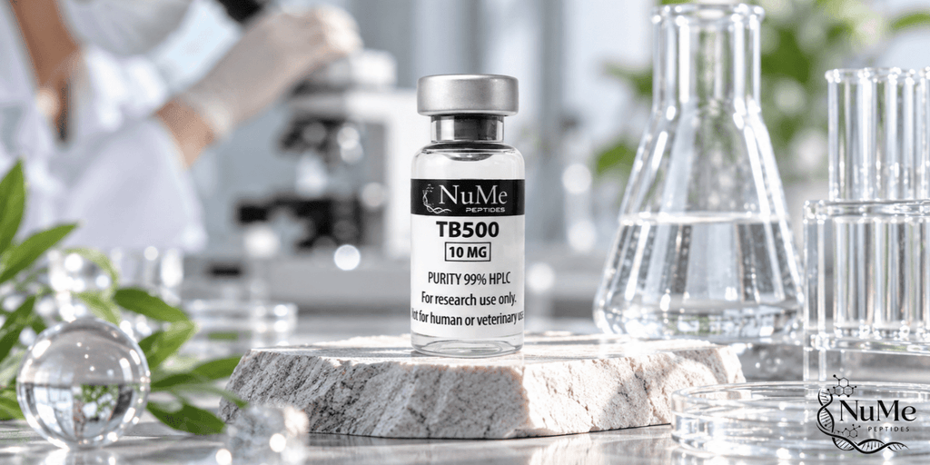
Explore batch-specific analytical details and research-use information for TB-500 (10 mg).
What Does Current Research Show About Peptides and Muscle Recovery?
Current research suggests that experimental peptides can influence several processes associated with skeletal-muscle regeneration. However, the evidence does not support treating mechanistic findings as established human muscle-recovery outcomes.
BPC-157 evidence remains mainly experimental
BPC-157 has shown repair-associated effects in multiple preclinical musculoskeletal studies.
Nevertheless, the evidence base remains dominated by animal research. The 2025 systematic review identified only one human study among 36 eligible publications. (Vasireddi et al., 2025)
Therefore, findings involving muscle structure, vascular signalling, or inflammatory pathways should be interpreted as experimental observations rather than established human benefits.
Thymosin β4 shows mechanistic activity
Thymosin β4 research provides evidence related to:
- myoblast migration;
- cytoskeletal regulation;
- tissue regeneration;
- vascular responses.
However, changes in regenerative markers do not automatically demonstrate restored muscle function.
This distinction was illustrated in an experimental mouse study in which thymosin β4 was associated with increased regenerating muscle fibres without a corresponding significant improvement in measured skeletal-muscle strength. (Thymosin β4 Muscle Function Study, 2010)
Functional endpoints are essential
Researchers should distinguish among three levels of evidence:
| Research outcome | What it indicates |
|---|---|
| Molecular signalling | A biological pathway has changed |
| Tissue regeneration | Structural repair may be occurring |
| Functional recovery | Muscle performance has improved |
An experimental compound may influence the first or second level without achieving the third.
For this reason, high-quality muscle-recovery research should ideally evaluate multiple endpoints rather than relying on one molecular marker.
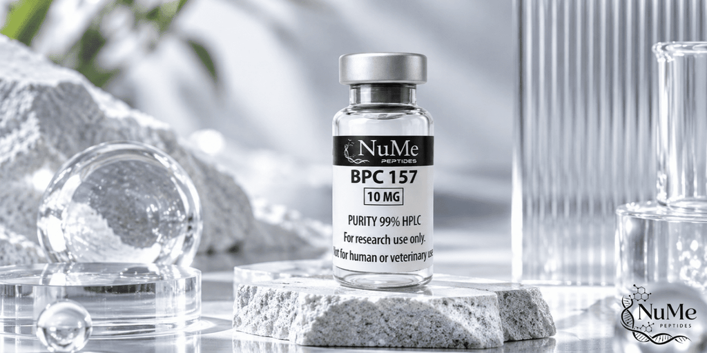
Explore batch-specific specifications, analytical documentation, and research-use details for BPC-157 (10 mg).
FAQs About Peptides for Muscle Recovery Research
What Are Peptides for Muscle Recovery Research?
They are peptide compounds investigated for potential effects on biological processes involved in skeletal-muscle regeneration, including inflammation, satellite-cell activity, myoblast migration, angiogenesis, and extracellular-matrix remodeling.
Is BPC-157 Proven to Improve Human Muscle Recovery?
No.
The current literature is dominated by preclinical studies, while robust human evidence remains limited. (Vasireddi et al., 2025)
Why Are Satellite Cells Important?
Satellite cells are resident skeletal-muscle stem cells.
Following injury, they can become activated, proliferate, differentiate, and contribute to repair or replacement of damaged muscle fibres.
Is TB-500 the Same as Thymosin β4?
They should not automatically be treated as identical.
Researchers need to confirm the molecular form, sequence, and analytical identity of the material used before applying findings from full-length thymosin β4 research.
Does More Muscle Regeneration Mean Greater Strength?
Not necessarily.
Histological regeneration and functional recovery represent different outcomes. Muscle strength also depends on fibre organization, fibrosis, neural function, connective tissue, and tissue mechanics.
Are BPC-157 and TB-500 Approved for Muscle Recovery in Canada?
Health Canada has identified BPC-157 and TB-500 among unauthorized injectable peptide products. Unauthorized products have not undergone Health Canada assessment for safety, efficacy, and quality. (Health Canada, 2026)
Conclusion
Research into peptides for muscle recovery research examines a complex biological system involving inflammation, satellite cells, myoblast migration, angiogenesis, extracellular-matrix remodeling, and muscle-fibre regeneration.
BPC-157 has generated substantial preclinical interest across musculoskeletal injury models, but human evidence remains limited. Thymosin β4 has mechanistic research involving actin regulation, cell migration, and skeletal-muscle regeneration, while TB-500 should be interpreted separately unless molecular identity has been clearly established.
Most importantly, researchers should distinguish molecular activity from tissue regeneration and tissue regeneration from functional recovery. A change in signalling, vascularization, or regenerating-fibre number does not independently prove restoration of muscle strength or clinical benefit.
For researchers looking for peptide materials for laboratory investigation, visit the Nume Peptides to explore the available research peptide collection.
Disclaimer: This content is provided for educational and scientific research purposes only. Research peptides are intended for laboratory research only and are not intended for human consumption or medical use.