
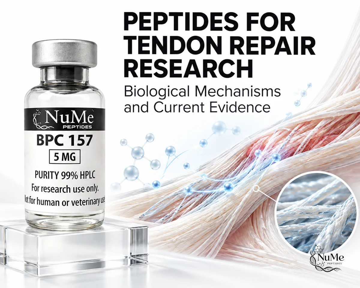
Peptides for Tendon Repair Research: Biological Mechanisms and Current Evidence


Research into peptides for tendon repair research examines how experimental peptide compounds may influence inflammation, tenocyte activity, collagen organization, vascular responses, extracellular-matrix remodeling, and mechanical recovery after tendon injury.
Tendons have a relatively limited intrinsic healing capacity compared with highly vascularized tissues. Their dense collagen structure, specialized cellular population, and comparatively low vascularity can make regeneration slow and may result in repaired tissue that differs structurally from uninjured tendon. Consequently, researchers are investigating biological approaches that may influence specific stages of tendon healing. (Docheva et al., 2015)
However, changes in a molecular pathway or histological marker should not automatically be interpreted as restored tendon function. Research must distinguish cellular activity, collagen remodeling, tissue organization, and biomechanical performance.
Canadian regulatory context is also relevant. Health Canada has identified BPC-157 and TB-500 among unauthorized injectable peptide products and states that unauthorized products have not been assessed for safety, efficacy, and quality in Canada. (Health Canada, 2026)
For researchers looking for peptide materials for laboratory investigation, visit the Nume Peptides to explore the available research peptide collection.
How Does Tendon Repair Occur After Injury?
Tendon repair involves overlapping inflammatory, proliferative, and remodeling processes. The objective is not simply to fill damaged tissue but to rebuild an organized collagen structure capable of transmitting mechanical force.
Inflammatory phase
Immediately after tendon injury, damaged vessels and cells initiate inflammatory signalling. Platelets, neutrophils, macrophages, and other immune components contribute to debris removal and release signalling molecules that influence later repair.
Researchers may evaluate:
- inflammatory cytokines;
- macrophage recruitment;
- growth-factor signalling;
- vascular responses;
- early extracellular-matrix changes.
Inflammation is necessary for repair, but excessive or prolonged inflammatory activity can contribute to disorganized matrix formation.
Proliferative phase
Fibroblast-like tendon cells increase their activity and contribute new extracellular matrix.
During this period, researchers may observe:
- increased cell proliferation;
- collagen production;
- temporary matrix formation;
- angiogenesis;
- migration of repair-associated cells.
Early repair tissue often contains a greater proportion of type III collagen compared with mature tendon.
Remodeling phase
Over time, the extracellular matrix undergoes gradual reorganization. Collagen fibres become more aligned with mechanical loading, matrix turnover changes, and type I collagen becomes increasingly important to the mature tendon structure.
Tendon healing can take months, and the resulting tissue may not fully regain the composition or mechanical properties of the original tendon. (Docheva et al., 2015)
| Tendon-healing stage | Main biological activity | Common research endpoint |
|---|---|---|
| Inflammation | Immune-cell recruitment and signalling | Cytokines and inflammatory cells |
| Proliferation | Cell activity and matrix deposition | Collagen and cell density |
| Early remodeling | Matrix reorganization | Collagen I/III balance |
| Late remodeling | Fibre alignment and maturation | Tensile strength and stiffness |
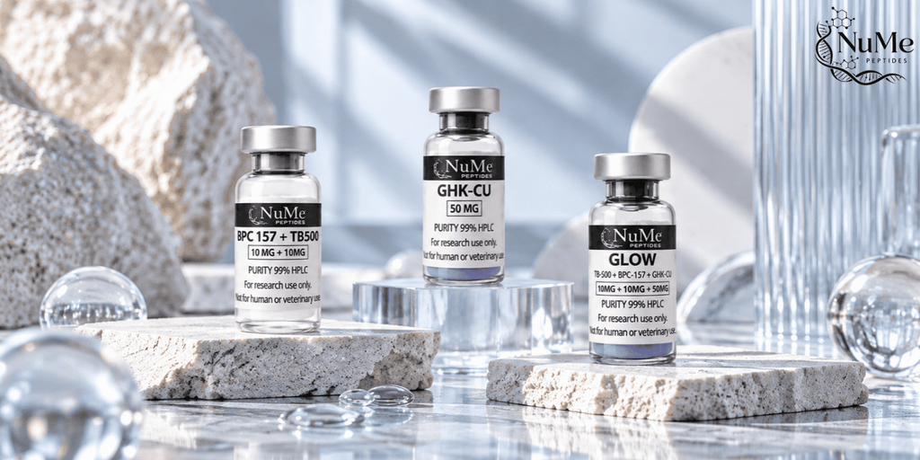
Explore research compounds studied for tissue repair, recovery, collagen support, and regenerative science in the Healing & Recovery collection.
What Roles Do Tenocytes, Collagen and the Extracellular Matrix Play in Tendon Repair?
Tendon structure is dominated by extracellular matrix, particularly collagen. Tenocytes and tendon-derived progenitor cells help maintain and remodel this matrix.
Tenocytes
Tenocytes are specialized tendon cells responsible for maintaining extracellular-matrix homeostasis. Following injury, tendon-associated cells may change their proliferation, migration, and matrix-producing activity.
Research often evaluates:
- tenocyte proliferation;
- cell migration;
- expression of tendon-related genes;
- collagen production;
- responses to mechanical loading.
Increasing cell number alone does not necessarily produce better tendon repair. The cells must also generate appropriately organized matrix.
Collagen I and collagen III
Type I collagen is the dominant collagen form in healthy tendon and contributes substantially to tensile strength.
Type III collagen tends to increase during early healing. It can provide temporary structural support but forms a less mechanically optimized network than mature type I collagen. As remodeling progresses, researchers often examine changes in the relationship between collagen types I and III.
Extracellular-matrix remodeling
The tendon matrix includes collagen, proteoglycans, glycoproteins, and other structural components. Matrix metalloproteinases and their inhibitors participate in controlled degradation and reconstruction of this environment.
Therefore, tendon repair requires a balance between:
- matrix synthesis;
- matrix degradation;
- collagen alignment;
- cross-linking;
- mechanical adaptation.
Research into tendon therapies increasingly focuses on restoring organized matrix architecture rather than simply increasing collagen production. (Docheva et al., 2015)
How Are Peptides Investigated in Experimental Tendon Repair Models?
Studies of peptides for tendon repair research use multiple experimental models because tendon healing involves both biological and mechanical processes.
Cell-based models
Laboratory studies may use:
- primary tenocytes;
- tendon-derived stem or progenitor cells;
- fibroblast-like cells;
- endothelial cells;
- inflammatory-cell cultures.
Researchers can then investigate whether a peptide affects migration, proliferation, collagen expression, signalling pathways, or cellular responses to stress. These models are useful for identifying mechanisms but cannot reproduce the complete mechanical environment of a tendon.
Animal tendon models
Animal studies can evaluate repair at the tissue level.
Researchers may measure:
- tendon thickness;
- collagen organization;
- inflammatory infiltration;
- vascularity;
- histological healing scores;
- tensile strength;
- failure load;
- stiffness.
This distinction matters because an intervention could improve histological appearance without producing equivalent improvements in mechanical strength.
Mechanical testing
Biomechanical outcomes are particularly important in tendon research. A repaired tendon ultimately needs to transmit force. Therefore, experiments may measure maximum load before failure, stiffness, elasticity, or other mechanical properties.
For this reason, evidence based only on cellular or molecular markers should not be treated as equivalent to evidence showing restored tendon function.
Which Peptides Are Being Studied in Tendon Repair Research?
BPC-157 and thymosin β4/TB-500-related compounds are frequently discussed in experimental musculoskeletal research, but their evidence profiles are not equivalent.
BPC-157
BPC-157 is a synthetic pentadecapeptide investigated primarily in preclinical models.
Experimental research has explored BPC-157 in relation to:
- tendon injury;
- fibroblast migration;
- angiogenic signalling;
- inflammatory pathways;
- collagen organization;
- tendon-to-bone healing;
- biomechanical recovery.
A 2025 systematic review evaluating BPC-157 in orthopaedic and sports-medicine research identified 36 eligible studies. Thirty-five were preclinical, while only one involved humans. Tendon, ligament, muscle, and bone models were represented, demonstrating substantial experimental interest but very limited clinical evidence. (Vasireddi et al., 2025)
The review also highlighted the absence of sufficient clinical safety data. Therefore, BPC-157 findings in tendon models should be interpreted as preclinical evidence rather than proof of human tendon-repair effectiveness.
TB-500 and thymosin β4
TB-500 is commonly associated with thymosin β4, but researchers should distinguish commercial terminology from specifically characterized molecules used in scientific studies. Thymosin β4 is a naturally occurring 43-amino-acid peptide that interacts with G-actin.
Research involving Tβ4 has examined:
- cytoskeletal regulation;
- fibroblast migration;
- endothelial-cell migration;
- angiogenesis;
- inflammatory signalling;
- wound and connective-tissue repair.
Experimental work has also identified biologically active fragments derived from thymosin β4. (Philp et al., 2010)
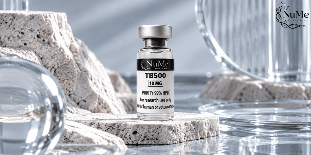
Explore batch-specific testing, analytical documentation, and research-use details for TB-500 (10 mg).
These mechanisms are relevant to tendon biology because cellular migration, vascular responses, and matrix organization contribute to repair. However, direct tendon-specific evidence for commercially described TB-500 remains substantially less established than the broader mechanistic literature surrounding thymosin β4.
Researchers should therefore confirm molecular identity before applying findings from full-length Tβ4 research to TB-500-labelled materials.
| Compound | Tendon-related research focus | Major evidence limitation |
|---|---|---|
| BPC-157 | Tendon healing, angiogenesis, fibroblast activity | Predominantly preclinical |
| Thymosin β4 | Migration, cytoskeleton, vascular signalling | Much evidence is mechanistic rather than tendon-specific |
| TB-500 | Tβ4-related research pathways | Cannot automatically be equated with full-length Tβ4 |
For experimental reproducibility, peptide sequence, lot information, HPLC data, mass-spectrometry results, and batch documentation should be considered when selecting research materials.
What Does Current Evidence Show About Peptides and Tendon Repair?
Current research suggests that experimental peptides can influence several biological processes relevant to tendon healing. However, the strongest evidence remains preclinical.
BPC-157 has experimental tendon evidence
Among commonly discussed experimental peptides, BPC-157 has been investigated relatively extensively in animal musculoskeletal models.
Studies summarized in the 2025 systematic review reported changes involving structural healing, angiogenesis, cellular responses, and biomechanical outcomes. Nevertheless, nearly all available studies were preclinical. (Vasireddi et al., 2025) This creates an important evidence gap between promising animal findings and demonstrated human outcomes.
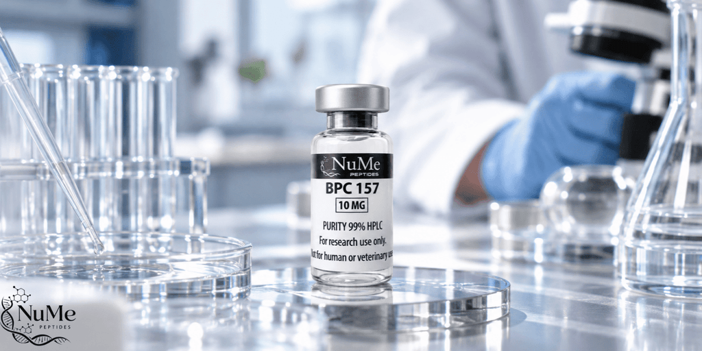
Explore batch-specific testing, analytical documentation, and research-use details for BPC-157 (10 mg).
Thymosin β4 evidence is more mechanistic
Thymosin β4 has biological evidence involving actin regulation, cell migration, vascular responses, and wound repair.
These mechanisms could be relevant to tendon healing, particularly during cell migration and early matrix reconstruction. However, evidence for direct tendon repair is less comprehensive than the wider Tβ4 literature.
Furthermore, TB-500 terminology should not be used interchangeably with full-length thymosin β4 unless molecular characterization confirms that relationship.
Tendon function requires more than faster healing
An experimental study may report increased collagen, greater cell migration, or altered angiogenesis. None of these measurements alone demonstrates complete tendon recovery.
Researchers should consider several levels of evidence:
| Research outcome | What it demonstrates |
|---|---|
| Molecular signalling | A biological pathway changed |
| Cellular response | Tenocyte or fibroblast behaviour changed |
| Histological repair | Tissue structure changed |
| Matrix organization | Collagen architecture improved |
| Biomechanical testing | Functional tissue properties changed |
The most convincing preclinical tendon research combines biological findings with structural and biomechanical measurements.
Canadian context
Health Canada has identified BPC-157 and TB-500 among unauthorized injectable peptide products in Canada. The agency states that unauthorized products have not been evaluated for safety, efficacy, and quality. (Health Canada, 2026)
This distinction is particularly important when interpreting peptides for tendon repair research: experimental biological evidence should not be presented as equivalent to therapeutic authorization.
FAQs About Peptides for Tendon Repair Research
What Are Peptides for Tendon Repair Research?
They are peptide compounds investigated for potential effects on biological processes involved in tendon healing, including inflammation, cellular migration, collagen production, angiogenesis, and extracellular-matrix remodeling.
Why Is Collagen Important in Tendon Healing?
Collagen provides most of the structural strength of tendon tissue. Type III collagen often increases during early repair, while remodeling gradually produces a more type-I-collagen-dominant and organized matrix.
Is BPC-157 Proven to Repair Human Tendons?
No. Current evidence is dominated by preclinical research. Human evidence remains insufficient to establish tendon-repair effectiveness or safety. (Vasireddi et al., 2025)
Is TB-500 the Same as Thymosin β4?
They should not automatically be treated as identical. Researchers should verify molecular sequence and identity before applying results from full-length thymosin β4 studies to TB-500-labelled research materials.
Does Increased Collagen Mean a Tendon Is Fully Repaired?
No. Collagen quantity, collagen type, fibre alignment, matrix organization, cross-linking, and mechanical strength all contribute to tendon function.
Are BPC-157 and TB-500 Approved for Tendon Repair in Canada?
Health Canada has identified BPC-157 and TB-500 among unauthorized injectable peptide products. Their appearance in experimental research should not be interpreted as Canadian therapeutic authorization. (Health Canada, 2026)
Conclusion
Research into peptides for tendon repair research examines a complex process involving inflammation, tenocytes, collagen production, extracellular-matrix remodeling, vascular responses, and restoration of biomechanical function.
BPC-157 has been investigated across several preclinical tendon and musculoskeletal models, but human evidence remains extremely limited. Thymosin β4 provides mechanistic evidence involving cell migration, actin regulation, and vascular signalling, while TB-500 should be interpreted carefully because commercial terminology does not necessarily establish molecular equivalence with full-length Tβ4.
Most importantly, researchers should distinguish biological signalling from structural healing and structural healing from functional tendon recovery. Increased collagen production or cell migration alone does not demonstrate restoration of normal tendon strength.
For researchers looking for peptide materials for laboratory investigation, visit the Nume Peptides to explore the available research peptide collection.
Disclaimer: This content is provided for educational and scientific research purposes only. Research peptides are intended for laboratory research only and are not intended for human consumption or medical use.